
Procedures
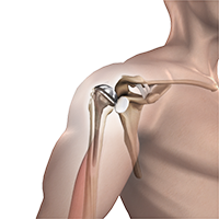
Anatomic and Reverse Total Shoulder Replacement
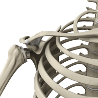
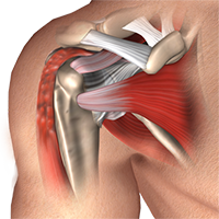
Arthritis can affect any joint in the body including the shoulder. All joints have cartilage on their surfaces including the glenohumeral joint which is the primary ball and socket joint of the shoulder. Cartilage is a smooth structure that covers the ends of the bones. In the shoulder, the top of the humerus or arm bone articulates with the glenoid cavity which is part of the scapula or shoulder blade.
Distal Clavicle Excision
A Mumford procedure or distal clavicle excision is a procedure performed for patients with arthritis at the acromioclavicular joint or distal clavicle osteolysis. The procedure is performed arthroscopically with removal of 5 to 6 mm of bone from the end of the clavicle to increase the space between the end of the collar bone and the opposite side of the joint (acromion). An open distal clavicle excision is also possible in cases of revision procedures or instability of the clavicle.
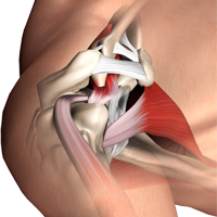
Rotator Cuff Repair
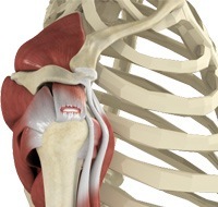
The rotator cuff is a group of tendons in the shoulder that provide support and enable a wide range of motion of the shoulder joint. Major injuries can cause rotator cuff tears. A rotator cuff tear is one of the most common causes of shoulder pain in middle-aged adults and older individuals.
Shoulder Arthroscopy
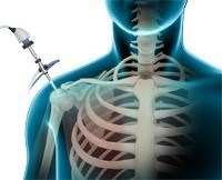
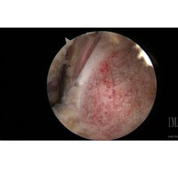
Arthroscopy is a minimally invasive diagnostic and surgical procedure performed for joint problems. Shoulder arthroscopy is performed using a pencil-sized instrument called an arthroscope. The arthroscope consists of a light system and camera to project images onto a computer screen for your surgeon to view the surgical site. Arthroscopy is used to treat disease conditions and injuries involving the bones, cartilage, tendons, ligaments, and muscles of the shoulder joint.
Shoulder Instability and Labral Repair
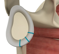
The glenohumeral joint of the shoulder, or the “ball and socket” joint, is purposely built to allow a wide range of motion while maintaining a stable relationship between the ball (humerus) and the socket (glenoid). The critical aspect of the shoulder is a balance between stability and motion. There are several structures that provide stability and failure of one or more of these restraints can lead to abnormal motion or instability.
Subacromial Decompression
Subacromial decompression surgery is performed to treat shoulder impingement, one of the most common causes of shoulder pain. Shoulder impingement causes the bones and tendons of the shoulder to rub painfully against each other when the arm is raised.
Revision Total Shoulder Arthroplasty
Total shoulder arthroplasty is the replacement of the head of the humerus (upper arm bone) and the glenoid cavity (cavity of the shoulder blade) into which the humerus fits, with artificial prostheses to relieve pain, swelling and stiffness caused due to damage of cartilage at the articulating surfaces.
Biceps Tenodesis
Proximal biceps tenodesis is the surgical reattachment of a torn proximal biceps tendon, which connects the upper part of your biceps muscle to the shoulder.
Suprascapular Nerve Release
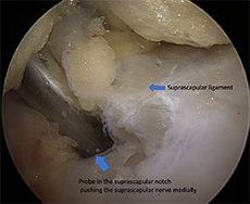
Suprascapular neuropathy can lead to significant shoulder pain and functional limitation. It occurs when the suprascapular nerve is injured along is course either due to compression or traction. Cysts in the suprascapular or spinoglenoid notches can compress the nerve.
Arthroscopic and Open Tendon Transfers
Tendon transfers are relatively uncommon procedures that can be utilized for either chronic nerve injury or irreparable tendon damage. Utilizing another muscle tendon unit to provide function for a deficient muscle can be very powerful in restoring function, strength and relieving pain in patients with chronic severe injuries.
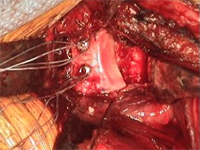
Open Instability Repairs
Shoulder instability is a chronic condition that causes frequent dislocations of the shoulder joint. A dislocation occurs when the end of the humerus (the ball portion) partially or completely dislocates from the glenoid (the socket portion) of the shoulder. A partial dislocation is referred to as a subluxation whereas a complete separation is referred to as a dislocation.
Latarjet Procedure
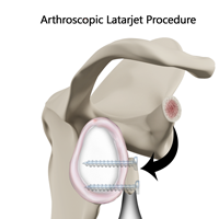
The shoulder joint provides a wide range of movement to the upper extremity, but overuse or trauma can cause instability to the joint. The Latarjet procedure is a surgical procedure performed to treat shoulder instability by relocating a piece of bone with an attached tendon to the shoulder joint.
Open Glenoid Bone Grafting
Open glenoid bone grafting is performed for patients with severe glenoid bone loss in the setting of recurrent anterior or posterior shoulder instability. For large glenoid bone loss (20% to 30%), treatment consists of a Latarjet procedure (coracoid transfer).
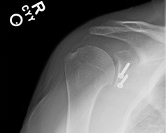
Open Reduction and Internal Fixation (ORIF)
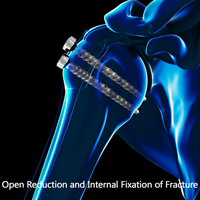
ORIF/open reduction and internal fixation is a surgical procedure employed for the treatment of a clavicle fracture not amenable to non-surgical conservative treatment.
Acromioclavicular Joint Reconstruction
The acromioclavicular (AC) joint is one of the joints present within your shoulder. It is formed between a bony projection at the top of the shoulder blade (acromion) and the outer end of the clavicle (collarbone). The joint is enclosed by a capsule and supported by ligaments.
Sternoclavicular Joint Reconstruction
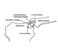
Sternoclavicular joint (SCJ) dislocations are uncommon and only account for 2 – 3% of all dislocations of the shoulder girdle. Most dislocations occur anteriorly while only 5 to 27% occur in a posterior direction. Anterior dislocations often do not require any acute treatment while posterior dislocations will require either closed reduction or surgical stabilization due to the proximity of vital structures posterior to the sternum.
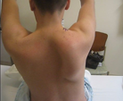
Arthroscopic Scapulothoracic Bursectomy
Scapulothoracic bursitis, or a "snapping" scapula, wasfirst described over 100 years ago as a condition with audibleand palpable grating localized to the superomedialangle of the scapula associated with pain. A variety of treatments have been described for patients with scapulothoracic bursitis including physical therapy, injections, open surgical resection of the superomedial angle of the scapula and bursectomy and arthroscopic scapulothoracic bursectomy.